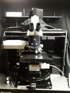
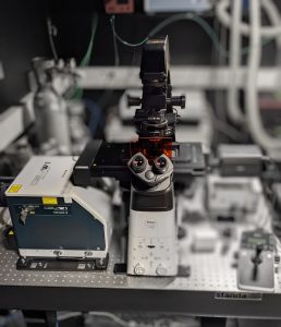
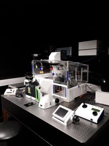
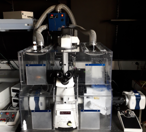
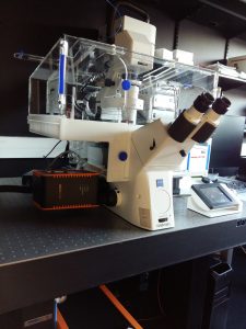
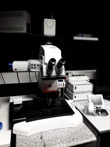
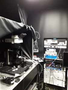
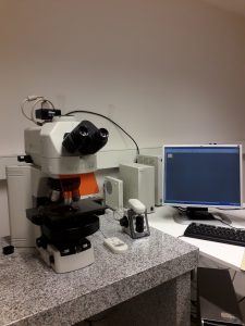
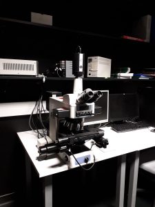
Microscopy became a prominent component of biological research. The Microscopy facility of the Orion technological core provide training, expertise and technical support for cell and tissue imaging, in vivo and in vitro, thanks to a large choice of microscope, covering a wide range of microscopy techniques.
To contact the microscopy facility, send an email to :
- All
- Confocal
- Widefield
- Spinning-disk
- Videomicroscope
- Multiphoton
- Lightsheet
Leica DMI6000 Upright TCS SP5 Confocal + Multi-photon (Room 307)
3D acquisition / Depth acquisition
Ex Vivo and fixed samples
Fixed stages : Motorized XY stage
Lasers :
– Argon 458, 477, 488, 496, 514 nm; 200mW
– DPSS laser 543 nm; 1.5 mW
– HeNe 633 nm; 15 mW
– Ti:Sapphire (Mai Tai®, Spectra-Physics) tunable from 690 to 1040 nm; 3W @ 800nm
Objectives :
– 10x/0.3 Dry WD: 11.0 mm
– 20x/0.7 Imm WD: 0.26-0.17 mm
– 40x/1.0 Oil WD: 0.08 mm
– 63x/1.4 Oil WD: 0.10 mm
– 25x/0.95 Water WD: 2,5 mm
– 63x/0.90 Water WD: 2.2 mm
Resonant Scanner for fast acquisition : 30 frames/sec at 512×512
Detectors :
– Confocal : 3 PMT and 2 HyD
– MP: 2 HyD NDD
Software : Leica LAS AF
Spinning-Disk Nikon CSU-W1 (Yokogawa) (Room 314)
Nikon Eclipse Ti-2 inverted microscope
3D acquisition of live or fixed samples / Fast imaging / Phase imaging / Dry-mass measurement
Cameras : sCMOS Hamamatsu 2304*2304/ pixel size : 6.5*6.5 µm; SID4 Phase camera (Phasics)
Motorized XY stage for multi-positions acquisition / Auto-Focus
Lasers:
– 405 nm; 150mW
– 488 nm; 100mW
– 561 nm; 100mW
– 638 nm; 100mW
Emission filters:
– BP 447/60
– BP 525/50
– BP 600/52
– BP 708/75
Objectives :
– Plan Fluor 10x/0.30 Dry WD: 16 mm
– Plan Apo lamD 20x/0.8 DIC Dry WD : 0.8mm
– Plan Fluor 40x/1.3 DIC Oil WD : 0.24mm
– Plan Apo lamD 60x/1.42 DIC Oil WD : 0.15mm
Software: NIS
Spinning-Disk Zeiss CSU-W1 (Yokogawa) (Room 315)
Zeiss Axioobserver Z1 inverted microscope
3D acquisition of live or fixed samples / Fast imaging
Environment chamber (temperature and C02 control)
Camera : sCMOS Hamamatsu 2048*2048 / pixel size : 6.45*6.45 µm
Motorized XY stage for multi-positions acquisition / Auto-Focus
Lasers:
– 405 nm; 100mW
– 445 nm; 80mW
– 491 nm; 150mW
– 561 nm; 100mW
– 642 nm; 100mW
Emission filters:
– BP 450/50
– BP 480/40
– BP 525/50
– BP 595/50
– LP 655
Objectives :
– 5x/0.16 Dry WD: 18.5mm
– 10x/0.45 Dry WD: 2.1mm
– 25x/0.80 LD LCI Plan-Apochromat DIC Imm WD: 0,57mm
– 40x/1.4 Plan-Apochromat DIC Oil WD: 0,13mm
– 63x/1.4 Plan-Apochromat PH3 Oil WD: 0,19mm
– 100x/1.4 Oil WD: 0.17mm
Software: Metamorph
Spinning-Disk CSU-X1 (Yokogawa) / TIRF (Room 314)
Nikon Eclipse Ti inverted microscope
3D acquisition of live or fixed samples / Fast imaging
TIRF microscopy
FRAP / FLIP
Environnement chamber (temperature and CO2 control)
Cameras :
– sCMOS Hamamatsu 2048*2048 / pixel size : 6.45*6.45 µm
– EM-CCD Evolve 512*512 / magnification lens 1.2 / pixel size : 13.3 µm*13.3µm
Lasers:
– 405 nm; 100mW
– 491 nm; 150mW
– 561 nm; 100mW
– 642 nm; 110mW
Emission filters:
– BP 445/45
– BP 525/39
– BP 605/64
– BP 725/150
– Quadband 440/40 – 521/20 – 607/34 – 700/45
Objectives :
– 4x/0.2 Dry WD: 20mm
– 10x/0.3 Dry WD: 16mm
– 20x/0.75 Dry WD: 1 mm
– 60x/1.4 Oil WD: 0.13mm
– 100x/1.4 Oil WD: 0.13mm
– 100x/1.49 Oil TIRF WD: 0.12mm
Software : Metamorph
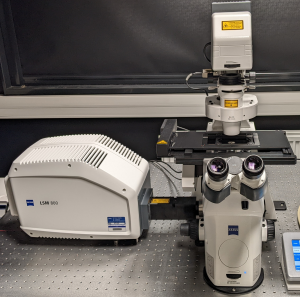
Zeiss LSM 800 Confocal
3D acquisition, Z-stack, time-lapse, spectral scan
Fixed samples
Motorized XY stage
Lasers :
– 405 nm ; 5mW
– 488nm ; 10mW
– 561 nm ; 10 mW
– 640 nm ; 5 mW
Objectives :
– 10x/0.30 Dry WD: 11mm
– 25x/0,80 IMM DIC WD:0,57mm
– 40x/1.4 Plan-Apochromat DIC Oil WD: 0.1mm
– 63x/1.4 Plan-Apochromat Oil WD: 0.1mm
Sensors: 2 Multialkali PMT and 1 spectral GaAsp detector
Software : ZEN
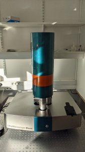
Ultramicroscope Blaze– LaVision Biotec (Room 305)
3D Fast image acquisitions of large samples
Living or cleared samples
Imaging solution : ECi ; CUBIC ; Water
Lasers:
– 488 nm ; 75mW
– 561 nm ; 75mW
– 640 nm ; 70mW
– 785 nm ; 75mW
Objectives :
– 1.3X/ 0.08 WD : 9mm
– 4X/ 0.3 WD : 6mm
(an additional lens provide a zoom ranging between 0,6x to 2,5x)
Camera : sCMOS pco.edge 5.5 2560*2160 / pixels size: 6.5 * 6.5µm
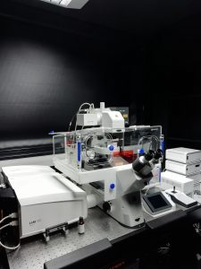
Zeiss LSM 980 Confocal – Airyscan 2
3D acquisition, Z-stack, time-lapse, spectral analysis, tiles, multi-positions
Fixed / Lived samples
Environment chamber for temperature and C02 control
Motorized XY stage + Auto-focus
Lasers :
– 405 nm ; 14 mW
– 445 nm ; 8 mw
– 488 nm ; 10 mw
– 514 nm ; 10 mW
– 561 nm ; 10 mW
– 639 nm ; 8 mW
Objectives :
– 10x/0.30 Dry PH1 WD: 5.2mm
– 20x/0.80 Dry WD: 0.55mm
– 25x/0.80 IMM DIC WD: 0.57mm
– 40x/1.3 Oil DIC WD: 0.21mm
– 63x/1.4 Oil DIC WD: 0.19mm
Sensors: 2 PMT, 1 PMT GasP spectral sensor , 1 Airyscan detector (v2.0)
Software : Zen blues
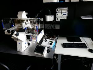
Zeiss AxioObserver 7 inverted – Videomicroscope (Room 318)
Widefield microscope
Fixed and lived samples
Motorized xyz stage + piezoelectric Z stage (150µm range)
Fluorescent lamp + white light
Environment chamber for temperature and CO² control
Objectives :
– 10x/0.45 Plan Apo Ph1 Dry WD: 2,1mm
– 20x/0.5 EC Plan-NeoFluar Ph2 Dry WD: 2mm
– 25x/0.80 LD LCI Plan-Apochromat DIC Imm WD: 0,57mm
– 25x/0.80 LCI Plan-Neofluar Ph2 Imm WD: 0,24mm
– 40x/1.4 Plan-Apochromat DIC Oil WD: 0,13mm
– 63x/1.4 Plan-Apochromat Oil WD: 0,19mm
Fluorescent filters sets :
– BP 433/25
– BP 482/25
– BP 520/35
– BP 555/25
– BP 600/38
– BP 647/55
– BP 680/40
Excitation central peak :
– 385 nm
– 430 nm
– 475 nm
– 511 nm
– 567 nm
– 630 nm
– 647 nm
Cameras : sCMOS (Hamamatsu Flash 4) 2048*2048 / pixel size : 6.45*6.45 µm
Autofocus capability (Definite Focus 2)
STEDYCON add-on
STEDYCON add-on
Additional module able to perform confocal and STED acquisition mounted on the Zeiss AxioObserver 7 Videomicroscope.
Scanning Frequency : up to 800 Hz
12 pinhole positions
4 Avalanche PhotoDiodes
Lasers:
– Continuous wave diode @405nm; 20mW
– Pulsed diode @488nm; 1,2mW @40MHz
– Pulsed diode @561nm; 0,2mW @40MHz
– Pulsed diode @640nm; 1,2mW @40MHz
– STED Laser @ 775nm; 1,25 W @40MHz
Software : Stedycon Smart Control
Objectives :
– 10x/0.45 Plan Apo Ph1 Dry
– 20x/0.5 EC Plan-NeoFluar Ph2 Dry
– 25x/0.80 LD LCI Plan-Apochromat DIC Imm
– 25x/0.80 LCI Plan-Neofluar Ph2 Imm
– 40x/1.4 Plan-Apochromat DIC Oil
– 63x/1.4 Plan-Apochromat Oil
– 100x/1.46 Plan-ApoChromat DIC Oil
Filter set :
– 450/60
– 525/40
– 600/50
– 675/50
Axio zoom V16 – Zeiss – Apotome.2 (Room 319)
Widefield or confocal-like observation of a wide range of sample sizes
Optical slices made possible with the Apotome structured illumination module
Very large field of view
Continuous zoom ability (16x-258x)
LED light source (X-CITE 110 LED)
Objective : 2.3x/0.57 Dry
Motorized XY stage for multi-positions acquisition
Camera : sCMOS (Hamamatsu Flash 4) 2048*2048 / pixel size : 6.45*6.45 µm
Fluorescent filter sets for blue, green, red and far-red stainings.
Software: Zen
Scientifica Two-photon Microscope (Room 307)
In vivo tissue and organism observation
2-photon excitation
Scientifica microscope stand and stage allowing great stability and space to work on small animals
Acquisition of 3D z-stack, time-lapse
Upright Scientifica microscope stand with 2 interchangeable object holders: – One for small animals
– One for brain slices
Laser: Ti:Sapphire (Mai Tai®, Spectra-Physics) tunable from 690 to 1040 nm; 3W @ 800nm
Resonnant scanner: 30 frames/sec at 512×512
Detectors: classic PMTs – 1 more sensitive GaAsP
Objectives:
– 4x/0.10 Dry WD: 19.0mm
– 10x/0.45 Dry WD: 4.0mm
– 16x/0.80 Water WD:0.3mm
– 20x/1.0 Water WD: 2.0mm
Powered by SciScan (Labview based solution)
Nikon 90i Eclipse upright – Widefield microscope (Room C2M.3)
Fixed sample observation
Motorized XY stage
Sources : Fluo Lamp
Fluorescent filter sets for bleu, cyan, green, red and fare-red stainings.
Objectives :
– 2x/0.06 Dry WD: 7.5mm
– 4x/0.13 Dry WD: 17.1mm
– 10x/0.45 Dry WD: 4mm
– 20x/0.75 Dry WD: 1mm
– 20x/0.75 IMM WD: 0.35mm
– 40x/1.3 Oil WD: 0.2mm
Camera: CCD color Nikon DS Ri1 12 bits / 1280*1024 / pixel size: 6.45*6.45
Software : NIS-Elements AR Version 3.10
Neurolucida station / StereoInvestigator (Room 319)
A powerful tool for creating and analyzing realistic, meaningful, and quantifiable neuron reconstructions from microscope images. Perform detailed morphometric analysis of neurons, such as quantifying:
– the number of dendrites, axons, nodes, synapses, and spines
– the length, width, and volume of dendrites and axons
– the area and volume of the soma
– the complexity and extension of neurons
A Stereo Investigator system for stereology gives you accurate, unbiased estimates of the number, length, area, and volume of cells or biological structures in a tissue specimen.
Nikon Eclipse E800 microscope
Fixed sample
Source : Fluo Lamp
Motorized XY stage
Objectives :
– 1x/0.04 Dry WD: 3.2mm
– 2x/0.1 Dry Wd: 8.5mm
– 4x/0.2 Dry WD: 15.7mm
– 10x/0.45 Dry WD: 4.0mm
– 20x/0.75 Dry WD: 1.0mm
– 40x/0.75 Dry WD : 0.2mm
– 60x/1.4 Oil WD: 0.21mm
Camera : CCD Qimaging Retiga 2000 Color / pixel size: 7.4 x 7.4 µm / 1600 x 1200 pixels
Managed by:


